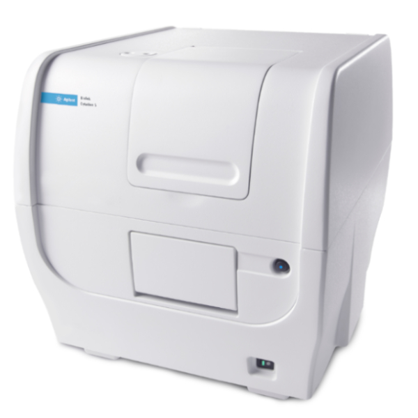

O Leitor Multidetecção com Captura de Imagens de Células Cytation™ 5 é a combinação de microscopia digital automática de campo amplo, com multidetecção convencional em microplacas, em um equipamento com design que permite upgrades. O módulo de microscopia oferece aumentos até 60x, em fluorescência, campo claro, campo claro de alto contraste, campo claro em cores e contraste de fase, permitindo o máximo em possibilidades de aplicações. Os módulos de multidetecção incluem detecção de fluorescência por filtros e monocromadores, luminescência e absorbância UV-Vis. O software Gen5™ fornece controle completo dos processos de captura de dados e de imagens, além de poderosas ferramentas de análise de dados e imagens.
Geral
| Modos de detecção | Absorbância UV-Vis Intensidade de fluorescência Luminescência Fluorescência polarizada Fluorescência resolvida no tempo Alpha |
| Métodos de leitura | Ponto final, cinetica, varredura de espectro, varredura de área do poço |
| Tipos de microplacas | Monocromadores: placas de 6 a 384 poços Filtros: placas de 6 a 1536 poços Captura de Imagens: placas de 6 a 1536 poços |
| Outros consumíveis compatíveis | Lâminas de microscopia, placas de Petri e de cultivo celular, frascos de cultivo celular (T25), câmara de contagem (hemocitômetro) Placas de Microvolume Take3 |
| Controle de temperatura | Incubação 4-Zone até 65 °C com Condensation Control; + 0,2 °C em 37 °C |
| Resfriamento | Módulo Opcional de Resfriamento Peltier mantém temperatura interna a < 1°C acima da temperatura ambiente. Permite rápido resfriamento após processos incubados. |
| Agitação | Linear, orbital e orbital dupla |
| Software | Gen5™ para Leitores de Microplacas e Sistemas de Imagens Gen5 Image+ e Image Prime disponíveis para análise completa de imagens Gen5 Secure para conformidade à 21 CFR Part 11 (opcional) |
| Automação | Compatível com BioStack e automação de outras marcas Compatível com Incubador Automático BioSpa 8 |
| Controle de CO2 e O2 (opcional) | Faixa: 0 – 20% (CO2); 1 – 19% (O2) Estão disponíveis modelos para ambos CO2 e O2 ou somente CO2 |
Sistema de Captura de Imagens
| Modo de Captura de Imagens | Fluorescência, campo claro, campo claro de alto contraste, campo claro em cores e contraste de Fase |
| Método para captura de imagens | Uma cor, múltiplas cores, Montagem, Lapso de Tempo, pilha Z |
| Processamento de imagens | Projeção Z, contraste de fase digital, “stitching” |
| Câmera | Sony CMOS de 16-bits, padrão ou amplo campo de visão |
| Capacidade de objetivas | 6 objetivas que podem ser trocadas pelo usuário |
| Objetivas disponíveis | 1.25x, 2.5x (efetivo 2.25x), 2.5x (efetivo 2.75x), 4x, 10x, 20x, 40x, 60x |
| Objetivas de contraste de fase disponíveis | 4x, 10x, 20x, 40x |
| Capacidade de cubos de filtros para captura de imagens | 4 cubos instalados ao mesmo tempo, que podem trocados pelo usuário, além do canal de campo claro |
| Cubos de filtros para captura de imagens disponíveis | DAPI, CFP, GFP, YFP, RFP, Texas Red, CY5, CY7, Laranja de Acridina, CFP-YFP FRET, Iodeto de propídio, clorofila, ficoeritrina, CY5.5, TagBFP, Alexa 568, Ex377/Em647 |
| Cubos LED para captura de imagens disponíveis | 365 nm, 390 nm, 465 nm, 505 nm, 523 nm, 590 nm, 623 nm, 655 nm, 740 nm |
| Funções automatizadas | Autofoco, intensidade dos LEDs automática, exposição automática |
| Método de foco automático | Autofoco baseado em imagem Autofoco treinado pelo usuário Autofoco por laser (opcional) |
| Controles de posição | Controle por software Controle por joystick (opcional) |
| Taxa de captura de imagem | Autofoco por imagens: 96 poços, 1 cor (DAPI), 4x, 6 minutos 96 poços, 3 cores, 4x, 12 minutosAutofoco por laser: 96 poços, 1 cor (DAPI), 4x, <3 minutos 96 poços, 3 cores, 4x, <7 minutos e 30 segundos Modo rápido: 10 fps, 1 poço, 1 cor com <=50ms de tempo de integração |
| Software de análise de imagens opcional | Gen5 Image+: Análise avançada de imagens Gen5 Image Prime: Análise avançada de imagens Gen5 Secure Image+: Análise avançada de imagens, recursos para 21 CFR Part 11 |
Intensidade de Fluorescência
| Fonte de luz | Flash de xenônio |
| Detector | PMT para sistema de monocromadores PMT para sistema de filtros |
| Seleção de comprimentos de onda | Monocromadores quádruplos (superior/inferior) Filtros (superior) |
| Faixa de comprimentos de onda | Monocromadores: 250 – 700 nm (opcional até 850 nm) Filtros: 200 – 700 nm (opcional até 850 nm) |
| Banda de passagem do monocromador | Variável, de 9 nm até 50 nm em incrementos de 1 nm |
| Faixa dinâmica | 7 décadas |
| Sensibilidade | Filtros: 0,25 pM de fluoresceína (0,025 fmol/poço, placa de 384 poços)Monocromadores quádruplos: 2,5 pM de fluoresceína (0,25 fmol/poço, placa de 384 poços) – superior 4 pM de fluoresceína (0,4 fmol/poço, placa de 384 poços) – inferior |
| Velocidade de leitura (cinética) | 96 poços: 11 segundos 384 poços: 22 segundos |
Luminescência
| Faixa de comprimentos de onda | 300 – 700 nm |
| Faixa dinâmica | >6 décadas |
| Sensibilidade | Monocromadores: 20 amol ATP (flash) Filtros: 10 amol ATP (flash), 100 amol (glow) |
| Faixa de comprimentos de onda | 300 – 700 nm |
| Faixa dinâmica | >6 décadas |
| Sensibilidade | Monocromadores: 20 amol ATP (flash) Filtros: 10 amol ATP (flash), 100 amol (glow) |
Fluorescência Polarizada
| Fonte de luz | Flash de xenônio |
| Detector | PMT |
| Seleção de comprimento de onda | Filtros |
| Faixa de comprimentos de onda | 280 – 700 nm (opcional até 850 nm) |
| Sensibilidade | Desvio padrão de 1,2 mP em 1 nM de fluoresceína |
| Fonte de luz | Flash de xenônio |
| Detector | PMT |
| Seleção de comprimento de onda | Filtros |
| Faixa de comprimentos de onda | 280 – 700 nm (opcional até 850 nm) |
| Sensibilidade | Desvio padrão de 1,2 mP em 1 nM de fluoresceína |
Fluorescência Resolvida no Tempo
| Fonte de luz | Flash de xenônio |
| Detector | PMT |
| Seleção de comprimento de onda | Monocromadores quádruplos (modo secundário) Filtros (superior) |
| Faixa de comprimentos de onda | Filtros: 200 – 700 nm (opcional até 850 nm) |
| Sensibilidade | Filtros: 40 fM de Európio (4 amol/poço, placa de 384 poços) Monocromadores: 1200 fM de Európio (120 amol/poço, placa de 384 poços) |
Absorbância
| Fonte de luz | Flash de xenônio |
| Detector | fotodiodo |
| Seleção de comprimentos de onda | Monocromador |
| Faixa de comprimentos de onda | 230 – 999 nm, em incrementos de 1 nm |
| Banda de passagem do monocromador | 4 nm (230 – 285 nm), 8 nm (>285 nm) |
| Faixa dinâmica | 0 – 4,0 DO |
| Resolução | 0,0001 OD |
| Correção de passo óptico | sim |
| Exatidão de comprimento de onda do monocromador | +2 nm |
| Repetibilidade de comprimento de onda do monocromador | +0,2 nm |
| Exatidão de DO | <1% em 2,0 DO <3% em 3,0 DO |
| Linearidade de DO | <1% de 0 a 3,0 DO |
| Repetibilidade de DO | <0,5% em 2,0 DO |
| Luz espúria | 0,03% em 230 nm |
| Velocidade de leitura (cinética) | 96 poços: 11 segundos 384 poços: 22 segundos |
Detecção Alpha
| Fonte de luz | Laser de 680 nm, 100 mW |
| Detector | PMT |
| Seleção de comprimento de onda | Filtros (superior) |
| Sensibilidade | 100 amol de peptídio LCK (placa de 384 poços de volume reduzido) |
| Fonte de luz | Laser de 680 nm, 100 mW |
| Detector | PMT |
| Seleção de comprimento de onda | Filtros (superior) |
| Sensibilidade | 100 amol de peptídio LCK (placa de 384 poços de volume reduzido) |
Injetores de reagentes (opcional)
| Modos de detecção suportados | Todos os modos |
| Número | 2 bombas seringas |
| Consumíveis compatíveis | Placas de 6 a 384 poços, placas de Petri e de cultivo |
| Volume morto | 1,1 mL com reversão de fluxo |
| Volume de dispensação | 5 – 1000 µL em incrementos de 1 µL |
| Tipos de placas | Microplacas de 6 a 384 poços, placas de Petri |
| Exatidão de dispensação | +1 µL ou 2% |
| Precisão de dispensação | <2% em 50 – 200 µL |
Características Físicas
| Potência | Consumo máximo de 250 Watts. |
| Dimensões | 50,8 cm P x 41,91 cm L x 44,5 cm A (20″ x 16.5″ x 17.5″) |
| Peso | 36,3 kg (80 lbs) |
| Potência | Consumo máximo de 250 Watts. |
| Dimensões | 50,8 cm P x 41,91 cm L x 44,5 cm A (20″ x 16.5″ x 17.5″) |
| Peso | 36,3 kg (80 lbs) |
Regulamentação
| Regulamentação | Com marcação CE e TUV. Em comformidade com RoHS. Estão disponíveis modelos para Diagnóstico In Vitro. |
Configurações
Configurações com Captura de Imagens e Multidetecção
| Número de Parte |
CYT5FV
|
CYT5FAV
|
CYT5MV
|
CYT5MPV
|
CYT5MFV
|
CYT5MFAV |
|---|---|---|---|---|---|---|
| CAPTURA DE IMAGENS | ||||||
|
Imagens por fluorescência
|
•
|
•
|
•
|
•
|
•
|
•
|
| Imagens por campo claro |
•
|
•
|
•
|
•
|
•
|
•
|
| Imagens por campo claro em cores |
•
|
•
|
•
|
•
|
•
|
•
|
| Imagens por contraste de fase |
•
|
|||||
| Baseado em FILTROS | ||||||
| Intensidade de fluorescência |
•
|
•
|
•
|
•
|
||
| Fluorescência polarizada |
•
|
•
|
•
|
•
|
||
| Fluorescência resolvida no tempo |
•
|
•
|
•
|
•
|
||
| Luminescência filtrada |
•
|
•
|
•
|
•
|
||
| Detecção Alpha |
•
|
•
|
||||
| Baseado em MONOCROMADOR | ||||||
| Absorbância UV-Vis |
•
|
•
|
•
|
•
|
||
| Intensidade de fluorescência |
•
|
•
|
•
|
•
|
||
| Fluorescência resolvida no tempo (secundário) |
•
|
•
|
•
|
•
|
||
| Luminescência |
•
|
•
|
•
|
•
|
||
| OUTROS | ||||||
| Compatível com placas Take3 |
•
|
•
|
•
|
•
|
Configurações apenas com captura de imagens ou apenas com multidetecção
| Número de parte |
CYT5V
|
CYT5PV
|
CYT5MF | CYT5MFA | CYT5F | CYT5FA | CYT5M |
|---|---|---|---|---|---|---|---|
| CAPTURA DE IMAGENS | |||||||
|
Imagens por fluorescência
|
•
|
•
|
|||||
| Imagens por campo claro |
•
|
•
|
|||||
| Imagens por campo claro em cores |
•
|
•
|
|||||
| Imagens por contraste de fase |
•
|
||||||
| Baseado em FILTROS | |||||||
| Intensidade de fluorescência |
•
|
•
|
•
|
•
|
|||
| Fluorescência polarizada |
•
|
•
|
•
|
•
|
|||
| Fluorescência resolvida no tempo |
•
|
•
|
•
|
•
|
|||
| Luminescência filtrada |
•
|
•
|
•
|
•
|
|||
| Detecção Alpha |
•
|
•
|
|||||
| Baseado em MONOCROMADOR | |||||||
| Absorbância UV-Vis |
•
|
•
|
•
|
||||
| Intensidade de fluorescência |
•
|
•
|
•
|
||||
| Fluorescência resolvida no tempo (secundário) |
•
|
•
|
•
|
||||
| Luminescência |
•
|
•
|
•
|
||||
| OUTROS | |||||||
| Compatível com placas Take3 |
•
|
•
|
•
|
Todas as configurações incluem incubação até 65 °C, agitação, e são compatíveis com o Módulo de Injetores de Reagentes, Controlador de Gases CO2/O2 e o Empilhador de Microplacas BioStack.