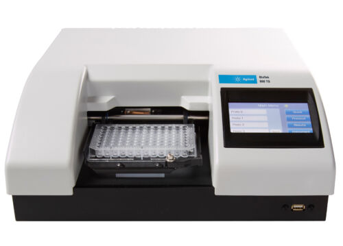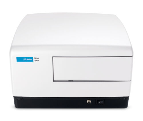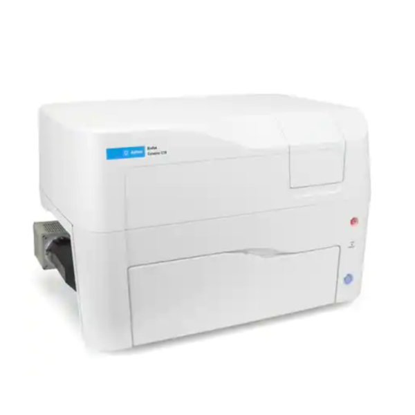

O Leitor com Captura Confocal de Imagens Cytation™ C10 combina microscopia automática digital confocal e de campo amplo, juntamente com leitura multimodal convencional de microplacas, tudo em um equipamento com design exclusivo e patenteado. O módulo confocal de disco giratório fornece excelente resolução e capacidades de seccionamento óptico em uma ampla variedade de tipos de amostras. Os componentes de alta qualidade utilizados no sistema, incluindo uma câmera CMOS científica (sCMOS) Hamamatsu, objetivas Olympus e iluminação por laser permitem excelente qualidade de imagens, a um preço significativamente mais baixo que outros equipamentos. O Cytation C10 conta ainda com ópticas de campo amplo para fluorescência, campo claro e contraste de fase. O módulo multimodal possui monocromador com banda de passagem variável, baseado no design já comprovado dos produtos Synergy da BioTek, líderes no mercado. Controles ambientais permitem ensaios com células vivas, e o software Gen5™ é especificamente projetado para tornar a detecção de amostras e captura de imagens um processo rápido e fácil. O novo visualizador 3D permite aos usuários reconstrução 3D de amostras mais espessas capturadas com microscopia confocal. O Cytation C10 traz microscopia confocal acessível a todos os laboratórios.
Geral
| Detection modes | UV-Vis absorbance Fluorescence intensity Luminescence |
| Read methods | Endpoint, kinetic, spectral scanning, well area scanning |
| Microplate types | Monochromator: 6- to 384-well plates Imaging: 6- to 1536-well plates |
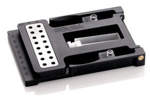
| Other labware supported | Microscope slides, Petri and cell culture dishes, cell culture flasks (T25), counting chambers (hemocytometer), Take3 Micro-volume plates |
| Temperature control | 4-Zone incubation to 45 °C with Condensation Control |
| Shaking | Linear, orbital, double-orbital |
| Software | Gen5 Microplate Reader and Imager Software included Gen5 Secure for 21 CFR Part 11 compliance (option) Gen5 Image+ and Image Prime software available for full image analysis (option) |
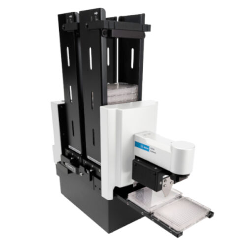
| Automation | BioStack and 3rd party automation compatible |
| CO2 and O2 control (option) | Range: 0 – 20% (CO2); 1 – 19% (O2), with optional Gas Controller Models for both CO2 and O2 or CO2 only are available |
Captura de imagens – Microscopia Confocal
| Imaging modes | Fluorescence |
| Image processing | Z-projection, digital phase contrast, stitching |
| Camera | Hamamatsu Orca sCMOS, 16-bit grayscale camera or Sony CMOS 16-bit grayscale camera |
| Objective capacity | 6-position automated turret for user-replaceable objectives |
| Objectives available | 20x, 40x, 60x |
| Image filter cube capacity | 4 user-replaceable fluorescence cubes |
| Imaging filter cubes available | CFP, CY5, DAPI, GFP, RFP, TRITC, brightfield |
| Laser | 6-line |
| Automated functions | Autofocus, user-trained autofocus, autoexposure, auto-LED intensity |
| Autofocus method | Image-based autofocus User-trained autofocus Laser autofocus (option) |
| Positional controls | Software Control Joystick controller (option) |
| Imaging methods | Single color, multi-color, time lapse, montage, z-stacking, z-stack montage |
| Field of view | Hamamatsu sCMOS: 0.65 mm + 5% at 20x magnification Sony CMOS: 0.70 mm + 5% at 20x magnification |
| Image collection rate | Laser autofocus, 0 ms delay, 96 wells: 8 mins, 9 secs Software autofocus, 0 ms delay, 96 wells: 12 mins, 1 sec |
Captura de imagens – Campo amplo
| Imaging mode | Fluorescence, phase contrast, color brightfield, user-selectable brightfield/high contrast brightfield |
| Imaging method | Single color, multi-color, time lapse, montage, z-stacking, z-stack montage |
| Image processing | Z-projection, digital phase contrast, stitching |
| Camera | Hamamatsu Orca sCMOS, 16-bit grayscale camera or Sony CMOS 16-bit grayscale camera |
| Objective capacity | 6-position automated turret for user-replaceable objectives |
| Objectives available | 1.25x, 2.5x (2.25x eff), 4x,10x, 20x, 40x, 60x |
| Phase objectives available | 4x, 10x, 20x, 40x |
| Image filter cube capacity | 4 user-replaceable fluorescence cubes, plus brightfield channel |
| Imaging filter cubes available | DAPI, CFP, GFP, YFP, RFP, Texas Red, CY5, CY7, Acridine Orange, CFP-FRET, CFP-YFP FRET, Chlorophyll, Phycoerythrin (PE), Propidium Iodide, CY5.5, TagBFP, Tag BFP-FRET, GFP (Ex)-CY5 (Em), RFP (Ex)-CY5 (Em), Alexa 568, Ex 377/Em 647, Oxidized roGFP2, TRITC |
| Imaging LED cubes available | 365 nm, 390 nm, 465 nm, 505 nm, 523 nm, 554 nm, 590 nm, 623 nm, 655 nm, 740 nm |
| Automated functions | Autofocus, autoexposure, auto-LED intensity |
| Autofocus method | Image-based autofocus User-trained autofocus Laser autofocus (option) |
| Positional controls | Software Control Joystick controller (option) |
| Image collection rate | Image-based autofocus: 96 wells, 1 color (DAPI), 4x, 6 minutes Laser autofocus: 96 wells, 1 color (DAPI), 4x, <3 minutes |
| Image Analysis Software option | Gen5 Image+: Image analysis Gen5 Image Prime: Advanced image analysis Gen5 Secure: 21 CFR Part 11 compliant features |
Intensidade de Fluorescência
| Light source | Xenon flash |
| Detector | PMT |
| Wavelength selection | Quad monochromators (top/bottom) |
| Wavelength range | 250 – 700 nm (900 nm option) |
| Monochromator bandwidth | Variable, from 9 nm to 50 nm in 1 nm increments |
| Dynamic range | 7 decades |
| Reading speed (kinetic) | 96 wells, sweep mode: 10 seconds 384 wells, sweep mode: 20 second |
Luminescência
| Wavelength range | 300 – 700 nm |
| Dynamic range | >6 decades |
Absorbância
| Light source | Xenon flash |
| Detector | Photodiode |
| Wavelength selection | Monochromator |
| Wavelength range | 230 – 999 nm, 1 nm increments |
| Monochromator bandwidth | 4 nm (230 – 285 nm), 8 nm (>285 nm) |
| Dynamic range | 0 – 4.0 OD |
| Resolution | 0.0001 OD |
| Pathlength correction | Yes |
| Monochromator wavelength accuracy | + 2 nm |
| Monochromator wavelength repeatability | + 0.2 nm |
| OD accuracy | <1% at 3.0 OD |
| OD linearity | <1% from 0 to 3.0 OD |
| OD repeatability | <0.5% at 2.0 OD |
| Stray light | 0.03% at 230 nm |
| Reading speed (kinetic) | 96 wells: 10 seconds 384 wells: 20 seconds |
Injetores de reagentes (opcional)
| Supported detection modes | All modes |
| Number | 2 syringe pumps |
| Supported labware | 6- to 384-well plates, Petri and cell culture dishes |
| Dead volume | 1.1 mL, with back flush |
| Dispense volume | 5 – 1000 µL in 1 µL increments |
| Plate geometry | 6- to 384-well microplates |
| Dispense accuracy | +1 µL or 2% |
| Dispense precision | < 2% at 50 – 200 µL |
Características Físicas
| Power | Instrument: External 250W (minimum), 24VDC power supply, compatible with 100-240 VAC @50-60Hz. Optional 6-channel laser light source: External 250W power supply, compatible with 100- 240VAC @ 50-60Hz. Optional Hamamatsu scientific camera: External 75W power supply, compatible with 100- 240VAC @ 50-60Hz. |
| Dimensions | 27″ W x 18.5″ H x 20″ D (68.6 cm x 45.72 cm x 50.8 cm) |
| Weight | 122 lbs (53.3 Kg) |
Regulamentação
| Regulatory | CE and TUV marked. RoHS Compliant. Models for In Vitro Diagnostic use are available. |
Configurações
| Número de parte | C10PHC2 | C10MPHC2 | C10PWC | C10MPWC | C10PW | C10MPW |
|---|---|---|---|---|---|---|
| Confocal (60 µm) Pos 1 |
•
|
•
|
•
|
• | ||
| Confocal (40 µm) Pos 2 |
•
|
•
|
||||
| Hamamatsu CMOS | • | • | ||||
| Câmera padrão |
•
|
•
|
• | • | ||
| Leitura multimodal |
•
|
•
|
• | |||
| Contraste de fase |
•
|
•
|
•
|
•
|
• | • |