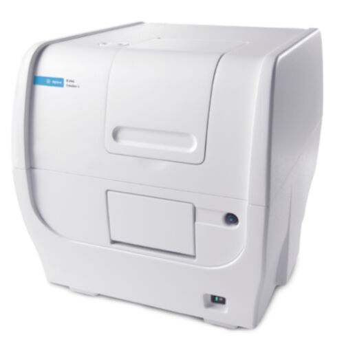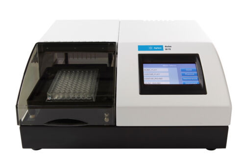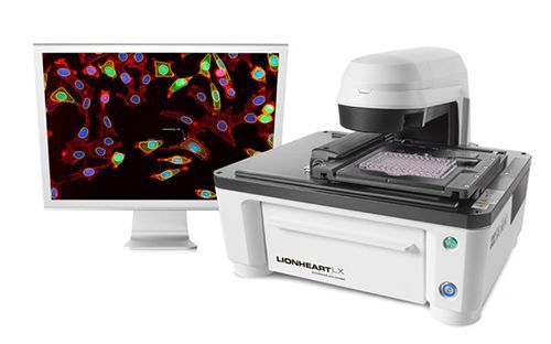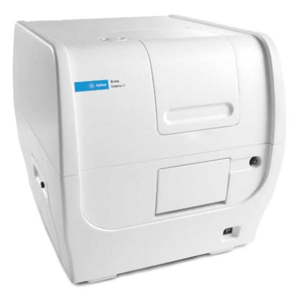

O Leitor Multidetecção com Captura de Imagens Celulares Cytation™ 7 combina microscopias digitais automáticas em modo superior vertical e invertida de campo amplo, com multidetecção convencional em microplacas, tudo em um desenho exclusivo e patenteado. O módulo de microscopia invertida permite visualização de amostras em aumentos desde 1.25x até 60x, por fluorescência, campo claro e campo claro em cores, para uma grande gama de aplicações. O módulo de microscopia superior vertical com luz refletida permite captura de imagens em aplicações como ELISpot ou varredura rápida de lâminas e detecção de regiões de interesse. O Cytation 7 possui monocromadores com banda de passagem variável, entregando o máximo em versatilidade e desempenho para leitura multidetecção de microplacas. Controle de temperatura até 45 °C, Módulo de Resfriamento Peltier opcional, controle de gases e agitação aumentam as possibilidades de aplicações em ensaios cinéticos com células. O Cytation 7 é controlado pelo software Gen5™, que combina poder de processamento e análise de imagens com a facilidade de uso.
Geral
| Detection modes | UV-Vis absorbance Fluorescence intensity Luminescence |
| Read methods | Endpoint, kinetic, spectral scanning, well area scanning |
| Microplate types | Monochromator: 6- to 384-well plates Imaging: 6- to 1536-well plates |
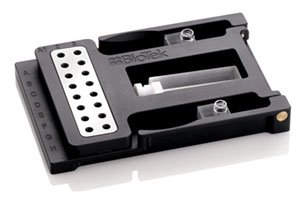
| Other labware supported | Microscope slides, Petri and cell culture dishes, cell culture flasks (T25), counting chambers (hemocytometer) Take3 Micro-Volume Plates |
| Temperature control | 4-Zone incubation to 45 °C with Condensation Control |
| Cooling | Optional Peltier Cooling Module maintains internal temperature with < 1 °C rise over ambient. Provides internal cooling after incubated processes. |
| Shaking | Linear, orbital, double orbital |
| Software | Gen5™ Microplate Reader and Imager Software included Gen5 Secure for 21 CFR Part 11 compliance (option) |
| Automation | BioStack and 3rd party automation compatible BioSpa 8 Automated Incubator compatible |
| CO2 and O2 control (option) | Range: 0 – 20% (CO2); 1 – 19% (O2), with optional Gas Controller Models for both CO2 and O2 or CO2 only are available |
Captura de imagens – Microscopia Invertida
| Imaging mode | Fluorescence, color brightfield, user-selectable brightfield/high contrast brightfield |
| Imaging method | Single color, multi-color, time lapse, montage, z-stacking, z-stack montage |
| Image processing | Z-projection, digital phase contrast, stitching |
| Camera | Sony CMOS, 16-bit grayscale |
| Objective capacity | 6-position automated turret for user-replaceable objectives |
| Objectives available | 1.25x, 2.5x (2.25x eff),2.5x (2.75x eff), 4x,10x, 20x, 40x, 60x |
| Image filter cube capacity | 4 user-replaceable fluorescence cubes, plus brightfield channel |
| Imaging filter cubes available | DAPI, CFP, GFP, YFP, RFP, Texas Red, CY5, CY7, Acridine Orange, CFP-FRET, CFP-YFP FRET, Chlorophyll, Phycoerythrin (PE), Propidium Iodide, CY5.5, TagBFP, Tag BFP-FRET, GFP (Ex)-CY5 (Em), RFP (Ex)-CY5 (Em), Alexa 568, Ex 377/Em 647, Oxidized roGFP2, TRITC |
| Imaging LED cubes available | 365 nm, 390 nm, 465 nm, 505 nm, 523 nm, 554 nm, 590 nm, 623 nm, 655 nm, 740 nm |
| Automated functions | Autofocus, autoexposure, auto-LED intensity |
| Autofocus method | Image-based autofocus User-trained autofocus Laser autofocus (option) |
| Positional controls | Software Control Joystick controller (option) |
| Image collection rate | Image-based autofocus: 96 wells, 1 color (DAPI), 4x, 6 minutesLaser autofocus: 96 wells, 1 color (DAPI), 4x, <3 minutes |
| Image Analysis Software option | Gen5 Image+: Image analysis Gen5 Image Prime: Advanced image analysis Gen5 Secure: 21 CFR Part 11 compliant features |
Captura de imagens – Microscopia Superior
| Imaging modes | Reflected color brightfield; transmitted color brightfield |
| Imaging methods | Single image, montage, time lapse, Z-stacking |
| Image processing | Z-projection, digital phase contrast, stitching |
| Camera | 16-bit Sony CMOS color camera |
| Lenses | Finder scope, 2x, 4x, 8x |
| Positional controls | Software Control Joystick controller (option) |
| Image collection rate | Entire 100 mm dish (montage) @ 1x: <3:00 minutes Entire microscope slide (montage) @ 1x: <1:15 minutes 96-well ELISpot plate @ 1x: <5 minutes |
| Image Analysis Software option | Gen5 Image+: Image analysis Gen5 Image Prime: Advanced image analysis Gen5 Secure: 21 CFR Part 11 compliant features |
Intensidade de Fluorescência
| Light source | Xenon flash |
| Detector | PMT |
| Wavelength selection | Quad monochromators (top/bottom) |
| Wavelength range | 250 – 700 (900 nm option) |
| Monochromator bandwidth | Variable, from 9 nm to 50 nm in 1 nm increments |
| Dynamic range | 7 decades |
| Sensitivity | Fluorescein 2.5 pM (0.25 fmol/well, 384-well plate) – top Fluorescein 4 pM (0.4 fmol/well, 384-well plate) – bottom |
| Reading speed (kinetic) | 96 wells: 11 seconds 384 wells: 22 seconds |
Luminescência
| Wavelength range | 300 – 700 nm |
| Dynamic range | >6 decades |
| Sensitivity | 20 amol ATP (flash) |
Absorbância
| Light source | Xenon flash |
| Detector | Photodiode |
| Wavelength selection | Monochromator |
| Wavelength range | 230 – 999 nm, 1 nm increment |
| Monochromator bandwidth | 4 nm (230 – 285 nm), 8 nm (>285 nm) |
| Dynamic range | 0 – 4.0 OD |
| Resolution | 0.0001 OD |
| Pathlength correction | Yes |
| Monochromator wavelength accuracy | +2 nm |
| Monochromator wavelength repeatability | +0.2 nm |
| OD accuracy | <1% at 3.0 OD |
| OD linearity | <1% from 0 to 3.0 OD |
| OD repeatability | <0.5% at 2.0 OD |
| Stray light | 0.03% at 230 nm |
| Reading speed (kinetic) | 96 wells: 11 seconds 384 wells: 22 seconds |
Injetores de reagentes (opcional)
| Number | 2 syringe pumps |
| Supported labware | 6- to 384-well plates, Petri and cell culture dishes |
| Dead volume | 1.1 mL with back flush |
| Dispense volume | 5 – 1000 µL in 1 µL increments |
| Plate geometry | 6- to 384-well microplates |
| Dispense accuracy | +1 µL or 2% |
| Dispense precision | <2% at 50 – 200 µL |
Características Físicas
| Power | 150 Watts maximum consumption |
| Dimensions | 16.4″ W x 17.5″ H x 20.2″ D (41.6 cm x 44.5 cm x 51.4 cm) |
| Weight | 80 lbs (36.3 kg) |
Regulamentação
| Regulatory | CE and TUV marked. RoHS Compliant. Models for In Vitro Diagnostic use are available. |
Configurações
| Número de Parte |
CYT7U
|
CYT7UW |
CYT7UMW
|
CYT7UM |
|---|---|---|---|---|
|
MICROSCOPIA
|
||||
| Microscópio vertical digital |
•
|
•
|
•
|
• |
| Microscópio invertido de campo amplo |
•
|
•
|
||
| Permite upgrade para microscopia invertida | • | • | ||
|
MULTIDETECÇÃO EM MICROPLACAS
|
||||
| Absorbância UV-Vis |
•
|
•
|
||
| Intensidade de Fluorescência |
•
|
•
|
||
| Luminescência |
•
|
•
|
||
| Permite upgrade para multidetecção |
•
|
•
|
||
|
OUTROS
|
||||
| Compatível com placa Take3 | • | • | ||
|
Incubaç
ão até 45 °C
|
• | • | • | • |